
A Caterpillar’s Life After Death
While I have written in the past about my interest in plant cell culture, my current project is with animals–an even more difficult group of cells to culture. Luckily, insect cells are much less finicky than mammalian cells, and methods have been established and refined since Thomas Grace at CSIRO first reported culturing cells from Opodiphthera eucalypti in 1962. During the ensuing five decades, advances were made in improving Grace’s media with endless variations, developing new formulas for sera-free defined media, and studying the effects of hormones, antibiotics, and additives on cell growth, differentiation, and protein synthesis. In addition, new protocols have been published for initiating cell lines from a growing breadth of terrestrial and aquatic arthropods and from a longer list of tissues, and for enlisting permanent cell lines in the production of insecticidal baculoviruses and transgenic proteins.
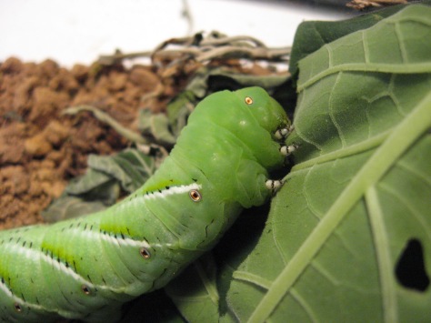
Despite all these advances, insect cell cultures are still Lepidoptera-centric, partly because many species are economically significant, well-characterized agricultural pests, and partly because they possess relatively large and easily reared larvae. These two attributes are especially apt in describing the tobacco hornworm (Manduca sexta), which for these very reasons has emerged as one of the de facto models for physiological, developmental, ecological, and behavioral studies of the insects as a whole and the Lepidoptera in particular. Although it thrives outdoors on solanaceous plants to the chagrin of home gardeners and tobacco farmers throughout the United States, captive populations maintained by scientists have readily adapted to crowded, sterile conditions on artificial diets. Larvae mature in just 2 or 3 weeks at a weight of 11 grams or more, allowing experiments to be conducted at nearly the same scale as would be for an adult lab mouse. And with the Manduca draft genome released just this August, we can expect many more insights from this big green worm.

But one way in which I have not seen Manduca fully utilized is in cell line development. Instead, the current focus in lepidopteran cell line research is less in mining various species for promising new lines and more in the development of protocols for transfecting insect cells to produce harvestable recombinant proteins for further study and pharmaceutical use. For these purposes, already well-entrenched cell lines like Sf9 from Spodoptera frugiperda and High-Five from Trichoplusia ni already suffice. M. sexta cell lines exist, but are limited to those from eggs and hatchling larvae. What I set out to accomplish with my project, then, was to start primary cell cultures from mature 5th instar Manduca larvae, with the overly-optimistic hope that one of these would later on establish a permanent cell line.

My target organs are larval midgut and fat body, which both contain their own stem cells, plus testes and wing imaginal discs that have a clear potential for growth as incipient adult structures. Last week, I dissected these organs and collected them in droplets of media, which I then held in sterile flasks and shut in my lab desk drawer for the weekend. Since this was all an exploratory study to prepare myself to compose a protocol and guide a class of students through it, I did not expect this to actually work.
But five days later:


The first picture shows a culture of numerous, very small cells having just migrated from an imaginal disc; the second picture directly above shows larger, more spread out cells from a fat body culture. Both cell types have adhered directly to the plastic; the organs themselves also attached over the last few days. Interestingly, while the fat bodies are still intact, the discs have completely disintegrated, leaving nothing but a naked tracheole framework and a puddle of small round cells busily secreting matrix. On the other hand, the fat body cells have assumed a fibroblast-like morphology, with a single clear organelle (possibly a nucleus?) visible. These were the only viable cell cultures thus far; the testes and midguts have not yet shown any activity so far.
I’m incredibly excited at the ease with which I was able to initiate these primary cultures, and it gives me hope that my fellow students will share in these beautiful results when I lead them through this experiment in a few weeks. After all, there is something exciting about taking cells from a complex, multicellular animal, then growing those cells in a flask indefinitely. In a way, it’s another way to keep the caterpillars that I’ve always been so fond of: Rather than nurturing individuals from the moment they are laid as eggs to their final shuddering wingbeats as worn, dying adults, I can remove their cells from that inevitable cycle of life and death and fix them in a state of eternal, fecund youth, stuck to the side of a plastic microcosm and swimming forever in nutrient-rich ambrosia. And on a moral level, I feel slightly absolved of my guilt of killing these magnificent animals for their cells: In a way, while their lives have been terminated, I’m giving their cells a shot at an immortal afterlife.
How Callus
Since graduation, I’ve been on a bit of a plant kick–not just because I needed to learn plant physiology and ecology for my GRE subject test and for my exploration of the flora and fauna near my new home near a suburban conservation area–but also because of my newfound appreciation for horticulture as art and discipline distinct from the botanical and ecological sciences, and for the therapeutic effect that gardening has had for my recent depression. As a side to my experimentation with tropical hibiscus propagation this summer, I was recently able to use my hibiscus calli in class in addition to the plant roots required for the day’s lab protocol. We performed assays to demonstrate areas of high enzymatic activity, and therefore identify zones of rapid cell growth and division, in these plant tissues. One of these was an assay for peroxidase, which neutralizes reactive oxygen species generated by metabolism that can destroy cell machinery and DNA.

Callus tissues are essentially stem cells in the somatic tissues of land plants. These stem cells are derived from more specialized cells that undergo rapid growth and division in the presence of growth hormones in the correct proportion. These callus form disorganized masses before differentiating into new roots and shoots, depending again on the appropriate mixture of hormones. In nature, callus tissue is encountered in plant wounds, where they grow and divide rapidly to heal over the injury. Calli are most frequently studied in vitro, however, since they are commonly used in plant tissue culture to produce numerous clones of a desired plant from single mature cells. In such situations, calli are the plant analogs of bacterial colonies in agar dishes.

I personally find it fascinating that such tissues can be generated on a hibiscus cutting in a jar of water, without sterile technique or specialized tools or media. Someday, I’d like to see if such in vivo callus cells are capable of forming plant embryos in culture, without having to induce calli in vitro. In fact, hibiscus cuttings are quite unique in how they develop such extensive masses of callus tissue all along the submerged part of the stem, not just in wounds. This disorganized tissue differentiates into functional roots in time, even without rooting hormone. This propensity to produce callus prior to rooting makes tropical hibiscus as a group rather easy to propagate, a trait that has made them so amenable to cultivation by enamored enthusiasts around the world. Soft green stem cuttings, taken any time of year, are relatively easy to root in water or soil, as long as adequate warmth and high humidity are provided.

My Wild Adventures With (Rooting) Datura Stem Cuttings
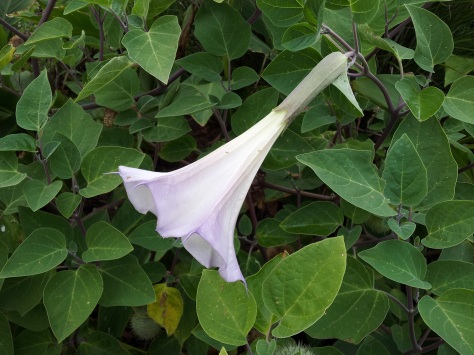
Taking a walk in Allegheny Commons a month ago, I noticed a weedy looking shrub peeking out amid the cannas and elephant ears flopping lazily under the wilting sun. Weedy, yes, but somehow it managed to fit in with its tropical ornamental neighbors. Its succulent purple stems and erect but bushy habit were attractive enough to catch my attention even next to the taller and flashier cannas. Velvety, dark green, heart-shaped leaves sheltered spiny seedpods nodding on thick stems from the forking shoot tips, like botanical maces melting away à la The Persistence of Memory. But these unusual features formed merely the backdrop to the huge, striking white blossoms, their scroll-shaped buds flushing a slight lavender as they unfurled into the late afternoon sky.
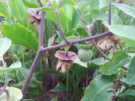
Anyone with an eye for gardening would recognize these exotic features anywhere on the planet as belonging to the plants of the genus Datura. Their common names are much more descriptive: jimsonweeds, thornapples, moonflowers, angel’s trumpets. Although their taxonomic history is rather complicated, I later tentatively identified this particular species as D. inoxia subsp. quinquecuspida, based on this old-ish online key from Erowid, adapted from one by Richard Sanders at the American Brugmansia and Datura Society (designated as the authority on Datura taxonomy by the ICNCP in 2004).
Datura‘s seductive pure white flowers and weedy abundance in out-of-the-way areas stand in eerily stark contrast to its storied reputation as an unpleasantly hallucinogenic drug and a deadly poison. I’ve seen Datura bushes in front of scruffy old homes in college neighborhoods (!) and cultivated in a row in front of suburban apartments filled with retirees (!!). Tom Scocca at the old Gawker website has a great essay in which he stumbles upon a Datura sprouting innocuously from a patch of bare dirt in the middle of New York City, blooming and reseeding and as wholly ignorant of its own toxic potential as all the daily passersby occupied with their own duties to grow and reproduce. Indeed, every afternoon when I would pick several D. inoxia flower buds to bloom at home, I would get strange looks from the dog walkers and outbound commuters who could not comprehend why I was so intently stomping around in an overgrown garden in an urban center.* But for me, the plant’s treacherous snake-in-the-grass character only augments the rare combination of grace and hardiness that convinced me that I had to have one of my own. Truly, for those who grow Datura on purpose (whatever purpose that might be), this is a plant that not only sits and looks pretty on its own, but has a dangerous reputation whose story adds to its mysterious, literally intoxicating charisma.

I took two D. inoxia cuttings on September 23 in the late afternoon, the worst season and time of day to take cuttings for most plants. Even worse, these were tip cuttings on a mature and blooming plant. For whatever reason (hmmm) there are not many definitive online guides on how to propagate Datura compared to its less infamous showy relative Brugmansia; many sources recommended propagation by roots or from seed, and only described stem cutting propagation for Brugmansia. However, these were stems I had used to feed my Manduca sexta hornworm caterpillar; after it died I only kept them around because I was too lazy to get rid of them. The whole time they sat neglected on my dresser, they remained succulent and green. The only change seemed to be the development of numerous small white bumps along the submerged sections of the stems. Meanwhile, the cutting wound appeared to turn brown and deteriorate.
Two weeks later, on October 6, I had to tidy up my room and noticed the first short root coming down out of the seemingly rotting wound on one cutting. By Saturday, October 8, that root had doubled in size and many more white tips were poking out of the wound; the other cutting had begun to root as well.

A closeup of the raised bumps on the cutting’s main stem.

Young roots growing from the wounds of two Datura cuttings. Note the extensive raised white bumps on the cutting on the right.

A closeup of the roots.
In retrospect, my success is not that surprising considering how related solanaceous plants are also commonly propagated by stem cuttings, like tomato plants and the aforementioned allied Brugmansia. In fact, I wonder if propensity to root by certain cutting techniques might be a way to differentiate similar populations or species. Certainly propensity to root is selected for under cultivation and some lines or varieties have higher success rates than others, so it probably has a genetic basis. It might even be a character state that develops and reverses in plant lineages; certain species might become better at rooting, like aquatic plants and succulents, or worse at rooting,like woody plants, as part of an adaptive syndrome to life in certain environments.
But TL;DR, here is how I would now recommend propagating Datura inoxia from stem cuttings:
- Get yourself some shoots from a flowering plant in late summer.
- Put them in water; leave alone for some weeks. No rooting hormone or any other cheats needed.
- When you’re done, now plant in suitable fast drying, low organic content soil and hope it overwinters in your house.
Not really complicated stuff, even if you’ve been hallucinating nightmares, your eyes are maximally dilated and you’re severely dehydrated, nauseous, dizzy, and just starting to separate dream from reality.

————————————————————————–
*One morning at the park, I did have an interesting conversation with a nice old lady who was picking the unripened alkaloid-laden fruits, which she claimed to extract in oil as a topical application for her back and leg pains. Hmm, OK, I said.

A Poem About Biological Systematics
Biological systematics is, let’s be honest… not a very sexy field of biology for most people. There is certainly something almost noble in the century-long quest to discover and classify all life on Earth and to unravel the deep and entangled thickets of the tree of life, to understand the literal origins of species. Not many scientific disciplines, I think, can boast such an inherent adventurousness. But that is all buried under layers of technical hemming and hawing about methods of tree construction, character validity, and taxonomic interpretation, all expressed in a jargon peculiar the the field, even within biology. So it’s not often that one sees systematics appear in the writings of non-scientists. But I did recently stumble on a poem, The Rose Family, by Robert Frost (1928):
The rose is a rose,
And was always a rose.
But the theory now goes
That the apple’s a rose,
And the pear is, and so’s
The plum, I suppose.
The dear only knows
What will next prove a rose.
You, of course, are a rose —
But were always a rose.
The poem lightly pokes fun at the seemingly pinheaded idea of apples, pears, and plums being classified in the same family, Rosaceae as regular roses. I’m sure Frost would have loved to know that blackberries and strawberries are in the Rosaceae as well; in fact, we now know that these two groups are even closer relatives of roses than Frost’s fruit trees.

Of course, the scientific foundation on which this poem rests is logically shaky (rose relatives are emphatically not roses, the same way your cousin, your relative, is not you) but the point is to explain one’s love as constant and steadfast in its devotion even as others’ attentions, tastes, and attitudes wander unpredictably over time. The use of the romantic connotation of flowers, in particular of the rose as opposed to less well-regarded blossoms, deepens the poem’s intentions. It’s a quintessential Frost poem: short, intense, and sweet, but executed with a light touch, like the fragrance of a rose.
(Featured image: An assortment of what most would call hawthorn (Crataegus), but are they actually roses?? From Wikipedia)
My First Animation
I spent a couple spare hours tonight fiddling with Animation Desk, a free basic drawing/animation app I found in the Microsoft app store. It’s really quite addictive, being flexible and easy to use even for those of us who have no art skills. I was so amused with my first effort that I just had to post it here:
(There are 12 frames in this video playing at 24 frames/second, making the whole thing only half a second long; if you play the video on Youtube you can change the settings in the lower right corner of the video to play at slower speed, or right click on the video to loop.)
I really got into animated television shows with Avatar: The Last Airbender in freshman year of college, and from there progressed onto anime as well as American-produced series. Animation has a reputation (in the U.S. at least) of being “for kids”, which often precludes them from being taken seriously by critics as works of art with messages as substantive as their live-action counterparts. If anything, one great advantage of animation is its enormous potential to transcend what is “real”, freeing an artist to exaggerate action, expressiveness, and dialogue as well as create worlds in which viewers can suspend all disbelief. Imagine Kill la Kill, a wild show with transforming school uniforms that examines how our clothes shape our identities and roles, without its schizophrenic transformation sequences and brilliant colors. Or what about The Venture Bros., a self-aware parody of popular science fiction and fantasy tropes, with its elaborate blunders and baroque machinations orchestrated not by its zany animated cast, but by live actors? And even for shows like Spongebob Squarepants, animation facilitates comedy by exaggerating facial expressions, distorting physics, and setting up scenarios whose impossibility in our world renders them all the more entertaining.
And lastly, animation is democratic. A complete, coherent episode may take skilled staff months to complete, but even so, any schmuck can, with a bit of time, stitch together a sequence of doodles into moving, living artwork. Hence this clip: 12 frames of a stick figure flying around with explosions, nothing compared to a complete episode or even a scene, but something I’m kind of proud of nonetheless.
Flies in Winter
This winter has been unusually mild, with snowfall not accumulating until late December and temperatures abnormally warm except for a few freezes in my area. There was one single huge snowstorm just a week ago that buried the northeast and the south, but that bypassed my area and was over quickly, even in places like NYC and Washington, DC that were hardest hit. And these past few days, we have been in the midst of a heat wave, with daytime temperatures reaching the 60s. Not so bad for mid-winter.
Because it was so sunny and warm yesterday, I decided to take some of the houseplants out in the backyard for some fresh air n’ photosynthesis. There, I made my first interesting natural history discovery of 2016.
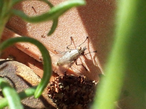
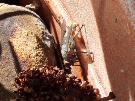
While moving my pelargoniums from the lawn onto the back porch, I noticed a pale insect huddling in the litter in the pot. This was a newly emerged fly, seen in the minutes after it emerged from its puparium, with its wings recently expanded but its cuticle still soft and unpigmented. How it ended up in the pot isn’t clear; I’m thinking it emerged from the lawn and climbed up and into the pot, rather than remaining dormant in the pot in my house throughout the winter. And these insects are relatively difficult to find in this state: many flies emerge, expand their wings, harden, and are ready for liftoff in as little as an hour.
You’ll notice this fly has a weird snout on its face. That snout is the remains of a structure called a ptilinum, a soft, bladderlike structure between the eyes which the fly repeatedly inflates with blood during its emergence. The puparium, the cramped outer shell that surrounds a fly pupa during its development, can be extremely tough to break from the inside, especially for an insect that has no biting mouthparts. But by pumping blood into its head and expanding the ptilinum, the fly uses a hydraulically powered battering ram to literally push the top off of the puparium and escape its time capsule. In some parasitic flies, the ptilinum is even used to break out of the earthen nests made by their bee hosts:
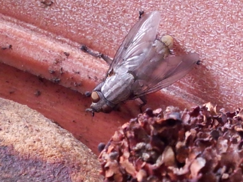
Given its importance in a brief but crucial time in the fly life cycle, the ptilinum is a pretty sophisticated structure, with associated musculature and sensory hairs. But once the fly has escaped, the ptilinum, now useless, begins to retract into the head. Over the next few days, the cuticle of the ptilinum dissolves, its muscles disappear, and all that is left externally is a thin line (the ptilinal suture) that marks the site where the ptilinum has shrunken.
Over the next thirty minutes, the fly colored up considerably. The stripes on the thorax indicate this fly is a flesh fly, in the large genus Sarcophaga. From these photos alone it is virtually impossible to pin the ID down to species.


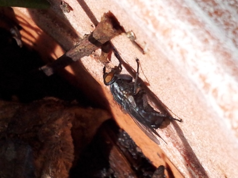
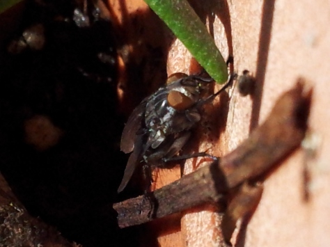
That afternoon I saw two other flies buzzing around outside; one came to settle on a plant dish for some water. Indeed, flesh flies, along with blow flies like Pollenia and Calliphora, are some of the earliest ‘large’ insects to appear outdoors, and many flies that overwinter as adults will come out briefly during warm sunny days and retreat when the cold returns. But I don’t know if it’s normal for flies like this one to break out of diapause in the middle of winter, or whether these callow individuals survive until spring without a chance to feed. But who knows whether this behavior is normal at all, what with the eerily uneventful winter we have this year?

Saturniid Hybrids
The giant silk moths in the family Saturniidae can be counted among the charismatic megafauna of the insects. Their richly patterned body coloration, sheer size, and exotic larvae make them popular among insect collectors, rearing hobbyists, and photographers, and they are even notable among the general public when they make their appearances to laypeople. Indeed, while relatively few in the scientific establishment seem to take the Saturniidae seriously as appropriate study organisms (a rant for another day), hobbyists in Europe and Asia, and to a lesser extent North America, have long bred and traded saturniids, observed and experimented with them informally, and occasionally documented their findings, sometimes in formal venues like low-profile peer-reviewed journals, but more often on personal websites, blogs, online fora, and other informal outlets.
One of the more intriguing aspects of the insect-keeping hobby I have been exploring lately is the hybridization of silk moth species. Bill Oehlke’s excellent website has a page that documents the outcomes of such experiments, many of which have had gorgeous results (check out the stunning moon moth hybrids involving Actias and the threatened Spanish moth Graellsia!). Many of these hybrid offspring are delicate creatures, making them doubly precious, and while many of these hybrids are undertaken by interested individual breeders, apparently there is a veritable European coalition of experienced and dedicated saturniid hobbyists that cross moths, distribute ova, and document each life stage of the hybrids as they develop.

On the surface, this all seems like a nice but perfectly useless Victorian gentlemen’s pastime, like collecting rare orchids or visiting exclusive gentleman’s clubs. However, saturniid hybridization experiments have yielded important scientific advances. The discovery by Walter Sweadner in the 1930s of a remote area in Idaho where two distinct populations of Hyalophora were interbreeding, for example, was the first to demonstrate that separate but related populations can hybridize and produce viable offspring in nature where their ranges overlap, even after they have already taken separate evolutionary paths. We now call these geographical areas ‘hybrid zones‘, and Sweadner’s studies helped established the foundations for our modern understanding of the role of interbreeding between populations during speciation. To this day, biologists continue to study hybridization in Hyalophora, a genus widespread in North America with regionally specialized but interbreeding populations.
Another hybrid, the oak tasar silk moth, Antheraea ‘proylei’, was developed as a multigenerational cross between domesticated A. pernyi males and wild A. roylei females in the 1970s, with the aim of boosting the Indian tussah silk industry in states with plentiful oak forest. Although the experiment was mainly a failure, A. ‘proylei’ is interesting from a genetic perspective because it is thought to be one of the few known natural cases of paternal mitochondrial DNA inheritance, with 98% of the mitogenome identical to A. pernyi (the males of which were backcrossed with the hybrids to found these lines). Also very unusual is that A. ‘proylei’ strains remain fertile indefinitely, despite mismatching chromosome numbers in the parent species (2n=98 for A. pernyi and 2n=62 for A. roylei), which is part of some convincing evidence that A. pernyi is merely a domesticated, genomically unstable descendant of A. roylei.

The whole point of this spiel was to introduce studies of giant silk moth hybrids as interesting, useful, and even foundational. I’ve been reading about these experiments because I want to attempt a little hybridization experiment of my own this summer, between my local strain of Antheraea polyphemus and a Japanese line of A. pernyi. Other hybrids have been produced among the unknown number of tropical and temperate Asian species, but these moths have all been rather same-y looking, reflecting the pretty uniform appearance of their parents. In the same vein, I have heard of hobbyists obtaining crosses of A. polyphemus with its close American relative A. oculea, and the resultant hybrid larvae and adults look almost exactly like regular A. polyphemus. Because the 4 New World species form a distant lineage separate from all their Old World cousins, and also because they differ in larval and adult characteristics, I am genuinely curious to see what hybrids of American and Asian species would look like, if they are viable at all.
Another reason why I’m interested in this project is because not many others have endeavored to do the same. Being that A. polyphemus is widespread in North America, A. pernyi is very common in Asian sericulture, and both have a history of use as study organisms in studies of insect development, diapause, and chemical communication, this is a cross that other hobbyists have surely already tried. However, I found fewer reports (at least, in English) on these attempts than I expected, and none described what interesting traits might be found in the offspring. The earliest attempt I could trace back was described in 1880 by an Alfred Wailly, who noted his lack of success:

On the other hand, Michael Collins & Robert Weast, in their 1961 book on the natural history of North American saturniids, state matter-of-factly that “Polyphemus freely crosses with [A. pernyi]. The larvae have been reared on oak.” And most notably, the Canadian lawyer Gary Botting, as a teenager in the late 1950s/early 1960s, carried out successful crosses* of Polyphemus moths (then named Telea polyphemus) with unspecified Asian moths (I assume either A. pernyi or A. yamamai), which supposedly convinced taxonomists to subsume the genus Telea as a North American branch of the genus Antheraea. (EDIT: see below)
*Per Wikipedia, because I have been completely unable to find the paper trail for this project or even verify the experiment’s importance in influencing taxonomic decisions. (EDIT: see below)
These few uninformative and disagreeing accounts don’t provide results, data, or even pictures for a cross with such evident potential, and I could find no evidence on my own that anyone has attempted to mate an A. polyphemus with a single A. pernyi since Botting’s science fair project in the 1960s. With this in mind, I have written this blog post as a sort of New Year’s resolution to myself to start this experiment this summer. In the coming months I will continue to research how to obtain hybrid pairings and find collaborators, and if I can get a generation of A. polyphemus larvae going this year I’ll be able to start! I’m definitely looking forward to this journey and its results!
EDIT (1/3/2016): I looked into Charles Michener’s 1952 monograph on the genera of the New World Saturniidae. Throughout the monograph, Michener discusses Antheraea as a genus, though he confusedly names it under a subgeneric level (probably a typo) when he actually gets to describing it. More importantly, he synonymizes Telea, that is, lists it as an invalid name with lower priority than the name Antheraea, and concludes:
 So even though Michener acknowledges that it was common practice at the time to use Telea polyphemus, he undoubtedly establishes Antheraea polyphemus as the correct name, nearly 10 years prior to Botting’s project.
How To Become a Test Prep Goon, Part 2
The first part of the application process was the simple application submission, phone interview, and the first checkpoint, the diagnostic exams, which I had outline in my previous post. Up to this point, everything had been conducted remotely by phone or email. However, now that I had passed the diagnostic exams, however, I was to come in for the face-to-face component of the process: the teaching audition, which would take place at the local company branch office.
The audition consists of a 5-10 minute presentation on a non-academic topic of the applicant’s choice, without using Powerpoint or other software, and with whiteboard and markers provided and props, as needed, brought by the applicant. This seems to be a standard prompt for the whole company as well as its competitors. Opinions on the Internet varied on how selective this step is compared to the diagnostic exams and training sessions, proper attire at the audition, and on various aspects of the presentation, such as the props, level of audience participation, and seriousness and complexity of the topic chosen.
I chose to talk about grafting, and specifically budding (the simplest of the myriad grafting techniques), due to my interest in gardening and plant biology. It’s a fairly dry-sounding topic compared to some of the other topics that people apparently have chosen (including falling in love, or crossing the street in Japan), but still original and interesting (compared to topics such as how to set up a chess board or play a sport). I’m also of the opinion that nothing needs to be intentionally gimmicky as long as it is conveyed with enthusiasm and backed up by extensive knowledge, and I felt sufficiently motivated by my own interest in plant biology and my gardening hobby to learn about the topic in depth. During the week I gave myself to prepare for my audition, I read online extension guides on budding, watched YouTube videos of gardeners demonstrating budding, and read relevant chapters from one recent, well-illustrated textbook on grafting. I also made sure to incorporate questions for the audience into my presentation (i.e., what might be some practical applications of grafting?), as well as a little dark humor.
The audition itself went relatively smoothly, since I was confident in my knowledge on the topic and had practiced a number to times to whittle my content down to 5-10 minutes. Some testimonials have said that auditions were done in front of the teaching staff and various other applicants, so I expected many more people and a lot more pressure, but this was not the case. The office was very small, and my audition took place in a tiny classroom in the back with only the very friendly regional coordinator and the course coordinator. I wore dress pants and a dress shirt for the occasion, which was surely still overdressing for this setting. Although I don’t remember the details of what I did during the audition (I tend to get selective amnesia during talks), I was able to exchange friendly banter with them for a few minutes and felt comfortable enough to ask for feedback on my performance, which seemed very positive. Though there were more applicants coming in to audition later on, I was told I had nothing to worry about.
Indeed, I was accepted to complete the next step this morning, sparing me a week of sitting around waiting to hear back, which was very considerate. At this point, after some paperwork, I would be moving on to the next step, being certified as a test prep instructor.
How to Become a Test Prep Goon, Part 1
Although I consider myself good at juggling multiple tasks when I need to, when I’m under no particular pressure I can’t help but fixate on a single goal at a time, especially if that goal is about to come to fruition in a matter of weeks. So during the past week, I’ve been working towards one lofty milestone: part-time employment!

Granted, I’ve been very picky about what full- and part-time jobs I’ve wanted to apply for, based mainly on how I can develop myself professionally. I’ve made the most progress in my applications as an MCAT instructor for a major test prep company. After going through one of the company’s MCAT courses and accomplishing the score I wanted to get on that exam, I was motivated, and felt I was qualified, to at least apply to teach two of the MCAT topics as an instructor with this company. Though I did hear that the pay is well above minimum wage for instructors of these classes, I cared more about just having something constructive and meaningful to fill my time for the next two years, and I think most individuals who apply to teach for these test prep companies feel the same way. So I’m going to detail my whole experience applying and interviewing for this position below.
I began my application process a few days after I had finished the GRE Biology subject test, since I now needed something else to occupy my time. The application process itself was simple, if a bit frustrating. The portal for applicants to search for positions and submit applications is very bare-bones and prone to glitches: the interface is ugly and difficult to navigate, the login page requires you to ask where you heard about the company EVERY. SINGLE TIME., and it doesn’t save your information so you have to complete your information in one sitting. I was astounded that the resume text extractor even worked correctly. However, once I submitted my first application (to be an SAT instructor), I completed my next two applications in quick succession with no trouble.
I moved on and forgot about all this until mid-November, when I was invited by email to have a phone interview with the regional coordinator/manager. The interview itself was very cordial and not at all what I expected, as it was more a confirmation of my teaching experiences and an overview of the extended interview, training process, and teaching responsibilities. Though the company had enough local SAT instructors, it had a few openings for MCAT instructors, so only my MCAT applications were really considered. I was told to expect to take two diagnostic exams, one for each MCAT subject for which I had applied to teach. If I passed these, I would next come to the company’s local outpost for a brief teaching audition. Once I passed these two hurdles, I would receive paid training for three days to qualify as an MCAT instructor with the company. It was implied that the interview was more of a notification that I was selected to continue through this extended application process than a weeding-out step in itself. However, it definitely helped to seek information on the Internet beforehand about the position, the pay, and the company’s culture, and I was able to use that information to take the initiative at the start of the interview and ask my questions first.
The two diagnostic exams were sent to me by email, and I was given several days to fill in my answers and submit them. I took this time to review my class notes from the course, then emailed my answers and scratch work back to the regional coordinator for grading. The same day I submitted them, I was notified that I had passed both exams, and I subsequently scheduled my teaching audition for this afternoon at their office.
